A patented surgical procedure for the treatment of scoliosis

Scoliosis surgery with pre-implantation CT scan and muscle preservation
The CCV Montpellier has created a unique collaborative process between radiologist and surgeon for the treatment of scoliosis

  • Optimal safety of implant placement

  • Back muscle preservation

  • Enhanced recovery after surgery

3 steps to rebalance the back

1 - CT scan guidance

The Interventional Radiologist

The procedure begins in the CT scan room, under anaesthesia, with the placement of pins to optimise implant trajectories.
The accuracy of the scan allows the radiologist to systematically maintain a safe distance from the spinal cord, even in the most severe deformities.

chir_scoliose

2 - Minimally invasive surgery

The surgical team

In the operating room, the marker pins will serve as a guide for placement of the screws, which were specifically developed at the CCV by Dr Thierry Marnay to preserve the muscles.
The rods will be inserted under the muscle to connect and align all the screws on the same side of the spine.
The paraspinal muscles will no longer need to be dissected as in conventional open surgery.

scoliose_vis

3 - Enhanced recovery after surgery

The Rehabilitation Centre

The muscle-sparing procedure with pre-implantation CT scan provides the best possible control of the adverse effects inherent in any operation.
A more anatomically correct approach means less blood loss, less post-operative pain and ultimately less anesthesia use.
All of these advantages are crucial for Enhanced Recovery After Surgery (ERAS) and for the patient to be up and about on the day of the operation.

recuperation

Spinal cord monitoring in the operating theatre

The CCV team is equipped with the Nim Eclipse device, dedicated to monitoring the spinal cord throughout the scoliosis correction procedure.
The Nim Eclipse device analyses the conduction of a very low amplitude current between the scalp and extremities of the limbs, thereby ensuring that the spinal cord is perfectly tolerant of spinal straightening manoeuvres.

Scoliosis SURGERY KEY POINTS

  • Indication: Scoliosis curvatures exceeding 40-50° at the end of puberty

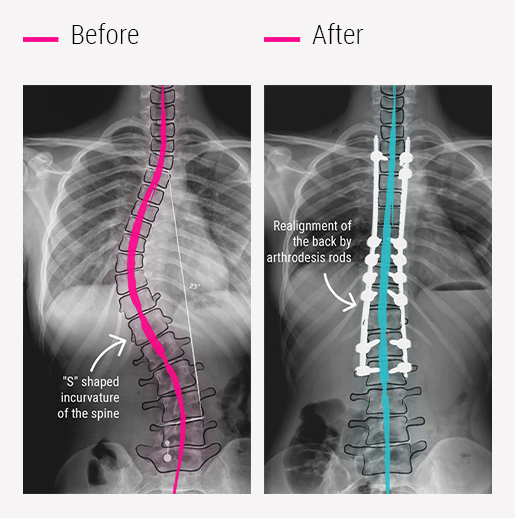
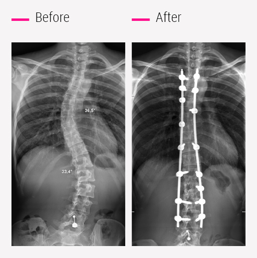
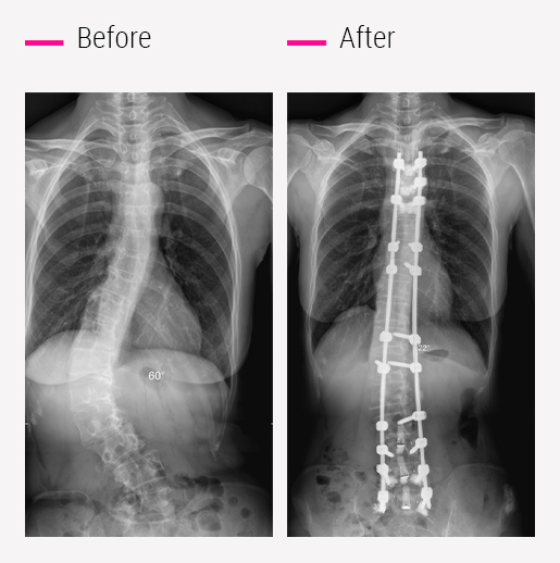
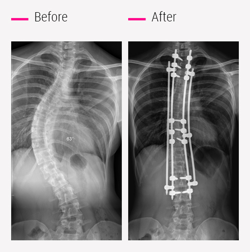
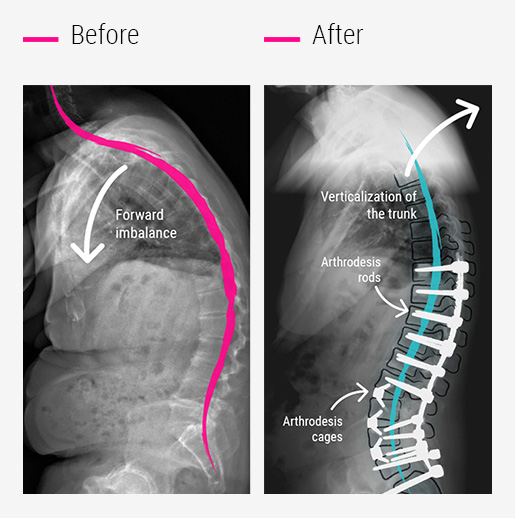
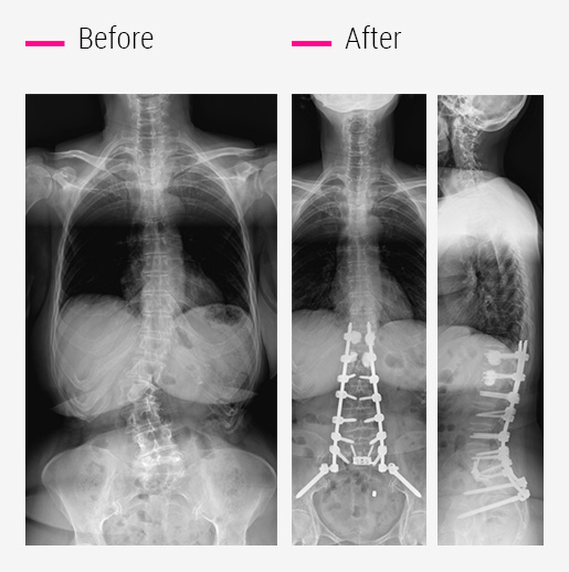
Minimally invasive and safe pre-implantation CT scan technique in the vertebrae

20 to 40 cm

Vertical scar in the back

1.30 to 4 hours

Duration of surgery 

4 to 7 days

Average length of hospital stay

6 weeks

Immobilisation with removable thermoformed brace (daytime)

3 to 9 months

Average recovery time

6 weeks

Minimum duration of school absence

3 months

Period before resuming sports activities

8th day

Flying home after surgery

6 to 12 months

Duration of evolution up to the final result of the surgery, (in the absence of neurological sequelae)

*These are mean times, with possibility of significant variability from one patient to another.

My E-Diagnosis

Wherever you are, access the services of an expert in spinal surgery

Request a free medical opinion, video teleconsultation, organise a customised medical stay… our e-medicine service offers you a whole range of services designed to give you access to a team of spinal surgery experts.

Find out more